Chester Knee Clinic News 2012
CKC Website - A Decade Online
2012 certainly promises to be a memorable year of celebrations. Here at Chester Knee Clinic, we have a landmark to celebrate too – our website been online for 10 years! The website was founded in 2002 and created with the help of Livewire Design and has been attracting a considerable amount of visitors ever since. We started kneeclinic.info with the aim of providing a one-stop online resource for information about Chester Knee Clinic's main areas of expertise, covering knee health, orthopaedics, sports injuries and rehabilitation, as well as a directory of specialists we work with.
Our website has grown to include specialised reference areas and knowledge bases, in particular on chondral and osteochondral injury and repair, and a place to catch up on all the latest news from our clinic and developments in the field of orthopaedics, imaging and physiotherapy.
We're constantly thinking about how to use the evolving digital landscape to improve and grow our resources, so look out for some exciting new features and updates coming to the site in the future.
CKC Research News
Updated on 10 November 2012
BioPoly™ RS Partial Resurfacing Knee Implant Clinical Trial
 This femoral
condyle partial resurfacing device is made from a microcomposite of Hyaluronic Acid and UHMWPE. This product is now available for use in the UK under
the terms of the clinical trial (multi-center, open label, prospective, consecutive series registry database of BioPoly™ RS partial resurfacing knee implant
clinical trial, approved by the NHS NRES Cambridge Central on 14 September 2011). This product has been CE marked for sale in the EU but it is not yet available for sale in
the US.
This femoral
condyle partial resurfacing device is made from a microcomposite of Hyaluronic Acid and UHMWPE. This product is now available for use in the UK under
the terms of the clinical trial (multi-center, open label, prospective, consecutive series registry database of BioPoly™ RS partial resurfacing knee implant
clinical trial, approved by the NHS NRES Cambridge Central on 14 September 2011). This product has been CE marked for sale in the EU but it is not yet available for sale in
the US.
We are now recruiting patients for this clinical trial (please see a list of Inclusion and Exclusion Criteria). Please be aware that we can not enter any potential candidates into the BioPoly clinical trial before they have been seen and examined in person at one of the sites by one of the investigators listed below. Email and telephone communication, however specific or detailed, are not sufficient for entry to this clinical trial.
Although the manufacturer of BioPoly implants will provide all implants free of charge for the duration of the clinical trial, your medical insurer or you will be responsible for all other costs (clinic appointments, imaging such as X-ray or MRI, the costs of the surgical procedure and hospital charges as well as perioperative physiotherapy). This does not apply if you enter the BioPoly clinical trial as an NHS patient.
The first BioPoly implantation was done early in January 2012 by Mr Dinesh Nathwani at the London Clinic (for more information please see the article from The Telegraph). The first large BioPoly (the "racetrack", picture above) was implanted in April 2012 by Mr Mike McNicholas, at the Warrington Hospital. Two BioPoly's were implanted in August and one more in October 2012 at Nuffield Health, the Grosvenor Hospital Chester. BioPoly Newsletter.
Participating Centres (in alphabetical order):
- Chester Knee Clinic at Chester Nuffield, contact: Mr Vladimir Bobic, office@kneeclinic.info
- London Bridge Hospital, contact: Mr Ian McDermot, london@sportsortho.co.uk
- The London Clinic, contact: Mr Dinesh Nathwani, d.nathwani@precisionsurgery.co.uk
- Warrington Hospital, contact: Mr Michael McNicholas, mike.mcnicholas@whh.nhs.uk
Further Information:
- BioPoly general information: www.biolpolyortho.com
- BioPoly video: http://youtu.be/MbzBSUz_82A
- Clinical Trials (a service of the US NIH): ClinicalTrials.gov (clinical trial identifier: NCT01473199)
- UKCTG - UK Clinical Trials Gateway (NHS National Institute for Health Research): BioPoly Registry Study
CKC Conference News
Updated on 12 October 2012
ACL Study Group Meeting
Jackson Hole, Wyoming, USA, 12 - 16 February 2012.
Please visit the ACL Study Group website for programme details and a list of members of the ACL Study Group.
BioPoly RS Knee Registry Study Investigator Meeting
Hilton Deansgate Hotel, Manchester (during the BOA meeting), 14 September 2012.
The next meeting of all investigators will be held in London in January 2013.
Nuffield Health GP Educational Seminar
Chester Race Course, 6 October 2012.
Chester Knee Clinic took part in this year's GP Educational Seminar (session on Winter Sport Injuries). Speakers: Vladimir Bobic, Andrew Phillipson, Mark Webb.
CKC Clinical News
Updated on 3 June 2012.
ACL Deficiency, Osteochondral Unit and OA
We will continue with our keen clinical interest and research in the aetiology, origins and pathways of post-traumatic osteoarthritis (or perhaps more correctly osteoarthrosis), which inevitably seems to follow many knee ligament injuries. We will try to consolidate the knowledge on the osteochondral unit (articular surface and subchondral bone) which is of paramount importance for understanding numerous factors which participate in the development of progressive accelerated knee wear and tear following a major knee trauma.
For example, a so-called bone bruise sustained during typical ACL injury is a series of trans-articular microfractures of variable severity which seem to recover well most of the time. However, this process also results in gradual and slow degeneration of articulating surfaces and subchondral bone. Many years later, neurovascular invasion at the osteochondral junction (sympathetic and sensory nerves are present within vascular channels in the articular cartilage) seems to be an important feature of both mild and severe OA (Source: Suri S et al, Ann Rheum Dis 2007;66:1423-1428).
It remains unclear why most initial lateral bone bruises recover well without clinically obvious lasting osteochondral damage, while medial articulating surfaces and subchondral bone deteriorate slowly over a long period of time (this process may take ten years or longer). The sensitivity of cartilage (and no doubt subchondral bone) to kinematic changes (a shift of the repetitive loading to a new location in ACL deficient knees) seems to initiate a slow degenerative pathway typically in the medial compartment (Source: Andriacchi TP et al., JBJSA 2009, 95;91 Suppl 1:95-101).
Please watch this space for further information on relevant articles and up-to-date knowledge on this controversial but extremely important topic.
Where does the OA pain come from?
Experimental and clinical observations suggest that the structural integrity of articular cartilage is dependent on normal subchondral bone turnover, intact chondrocyte function and ordinary biomechanical stresses. An increasing line of evidence suggests that there are strong communication and interaction between the subchondral bone and the articular cartilage. As bone and cartilage are closely interrelated, interventions affecting pain related to bone turnover may, in addition, be related to pain relevant to osteoarthritis. Pain is a central part of the clinical presentation of OA and the main reason for patients to seek consultation. Many and diverse structural features of the joint have been suggested to be involved in OA-associated pain, including, but not limited to, the presence of osteophytes in the patellofemoral compartment, focal or diffuse cartilaginous abnormalities, subchondral cysts, bone marrow oedema, subluxation of the meniscus, meniscal tears and Baker cysts. The literature also suggests an association of capsular distention with knee pain (an association of effusion with knee pain)
In athletes and younger adults who do not have osteoarthritis, traumatic knee injuries produce high-signal lesions in the medullary space extending to subcortical bone. These lesions are thought to represent contusions within the bone marrow and have been correlated with the occurrence of pain in athletes. Bone marrow lesions that are similar in appearance to those contusions have been noted in patients with knee osteoarthritis, but their association with the occurrence of pain in this disease is unknown.
Bone, and especially the subchondral bone plate, is involved in the pathogenesis of osteoarthritis. OA bone tissue is sclerotic yet undermineralized indicating abnormal bone cell metabolism. Studies in both human and animal models of OA support the concept that bone sclerosis could precede cartilage degradation and loss. Thus, subchondral bone sclerosis in OA may be due to abnormal osteoblasts characterized by increased metabolic activities that result in an increase in osteoid matrix that is undermineralized.
The correlation of bone marrow lesions with pain in knee OA has been convincingly established. Here, another compelling association is established between bone marrow (oedema) lesions and risk for progression of knee OA. What remains to be established is the cause-and-effect relationship between the various variables. For instance, whether an initial insult to articular cartilage leads to mechanical malalignment and subsequent subchondral bone destruction or rather subchondral bone damage leads to mechanical malalignment and subsequent articular cartilage destruction is not explained by the current analysis. In addition, the evolution and natural history of the baseline bone marrow lesions on follow-up MRI studies with respect to progression of knee OA has yet to be investigated. It is interesting to note that, histologically, the lesions that appear as bone marrow edema on MRI contain very little edema at all. Rather, they demonstrate fibrosis, osteonecrosis, and extensive bony remodeling and are likely the result of contusions and focal micro-fractures. These data contribute to a new understanding of the natural history of knee OA and suggests new areas of investigation and alternative treatment modalities to the few currently available.
Normal adult human articular cartilage is thought to be avascular and aneural. However, very recently, sympathetic and sensory nerves were both found to be present within vascular channels in articular cartilage in both mild and severe OA. Perivascular and free nerve fibres and nerve trunks have also been observed within the subchondral bone marrow and within the marrow cavities of osteophytes. Nerve endings localised in damaged human articular cartilage suggest that vascularisation and the associated innervation of articular cartilage may contribute to tibiofemoral pain in OA across a wide range of structural disease severity. The implications of these findings are that the musculoskeletal pain associated with osteoarthritis may result from a combination of the hitherto accepted central and bone-derived effects. A mechanism-based understanding of the factors involved in generating OA pain has gained acceptance and will further develop the area. Source: Lars Arendt-Nielsen, Hans Christian Hoeck: Peripheral and Central Sensitisation in Osteoarthritis – Implications for Treatment, European Musculoskeletal Review, 2011;6(3):158-61.
Recent Orthopaedic and Sport Injury News
Updated on 10 November 2012
A Randomized Trial Comparing Accelerated and Traditional Approaches to Postoperative Weightbearing Rehabilitation After Matrix-Induced Autologous Chondrocyte Implantation (MACI): Findings at 5 Years.
Jay R. Ebert, Michael Fallon, M.H. Zheng, David J. Wood, Timothy R. Ackland. Am J Sports Med July 2012 40 1527-1537.
Background: While structured postoperative rehabilitation after matrix-induced autologous chondrocyte implantation (MACI) is considered critical, very little has been made available on how best to progressively increase weightbearing and exercise after surgery.
Hypothesis: A significant improvement will exist in clinical and magnetic resonance imaging (MRI)–based scoring measures to 5 years after surgery. Furthermore, there will be no significant differences in outcomes in MACI patients at 5 years when comparing a traditional and an accelerated postoperative weightbearing regimen. Finally, patient demographics, cartilage defect parameters, and injury/surgery history will be associated with graft outcome.
Results: Of the 70 patients recruited, 63 (31 accelerated, 32 traditional) underwent clinical follow-up at 5 years; 58 (29 accelerated, 29 traditional) also underwent radiological assessment. A significant time effect (P < .05) was demonstrated for all clinical and MRI-based scores over the 5-year period. While the VAS demonstrated significantly less frequent pain at 5 years in the accelerated group, there were no other significant differences between the 2 groups. Between 24 months and 5 years, a significant improvement (P < .05) in both groups was observed for the sport and recreation subscale of the KOOS as well as a significant decrease (P < .05) in active knee extension for the traditional group. There were no significant differences (P > .05) in the MRI-based scores between 24 months and 5 years after surgery. Patient age and defect size exhibited significant negative correlations (P < .05) with several MRI-based outcomes at 5 years, while there were no significant correlations (P > .05) between clinical and MRI-based outcomes. At 5 years after surgery, 94% and 95% were satisfied with the ability of MACI to relieve their knee pain and improve their ability to undertake daily activities, respectively.
Conclusion: The outcomes of this randomized trial demonstrate a safe and effective accelerated rehabilitation protocol as well as a regimen that provides comparable, if not superior, clinical outcomes to patients throughout the postoperative timeline.
Other CKC News
Updated on 5 April 2012
Jackson Hole, Wyoming, USA
In February 2012, Chester Knee Clinic participated in an ACL Study Group meeting in Jackson Hole, a ski resort in the heart of the Grand Teton National Park in the north-western state of Wyoming. We were lucky enough to stay in Teton Village, located at the base of the mountain's aerial tram and gondola.
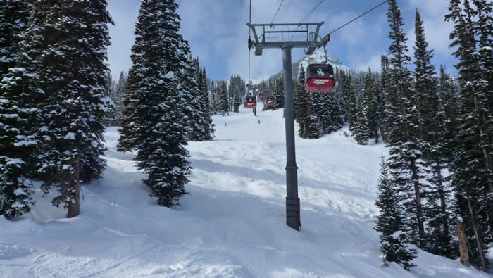
Jackson Hole is famous for miles of skiing in ungroomed natural bowls, plenty of high-altitude tree skiing and "Corbet's Couloir", a near-vertical 'corridor' runing between cliffs. Although Jackson Hole has a reputation as a tough mountain, we found it to be fantastic place with a great atmosphere, varied and challenging terrain and excellent snow conditions. We were very lucky as nightly snow showers provided a few inches of fresh powder almost every morning.
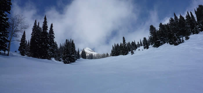
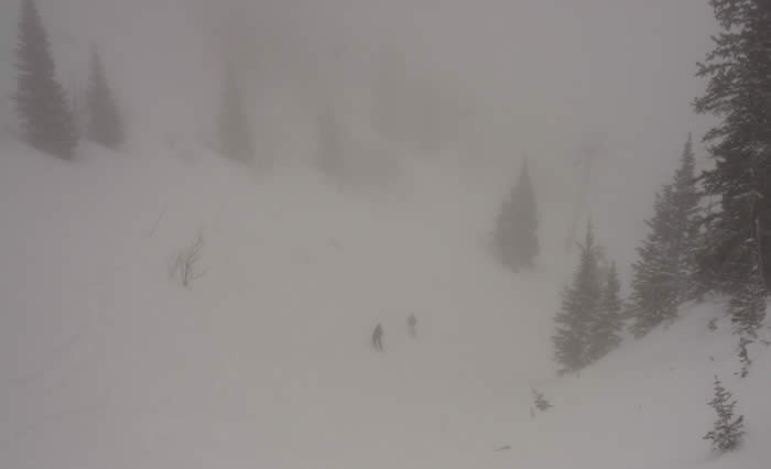
We particularly enjoyed skiing in large expanses of deep powder and in the woods, where we even spotted elk just a few yards away from us. With the help of experienced and knowledgable local ski guides, we made the most of the vast Rendezvous Mountain and all skied well. We all improved our skiing thanks to the challenging terrain and a certain long, natural half-pipe called "Dick's Ditch" (which was fun even in very limited visibility, as gravitational forces did the directional job). We are all back in one piece apart from a few bruises and minor neck and knee injuries.
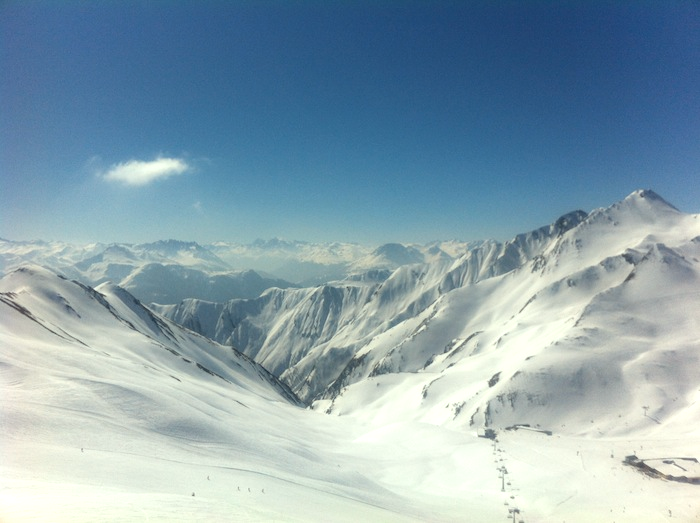
Kaunertal and Serfaus, Fiss & Ladis, Austria
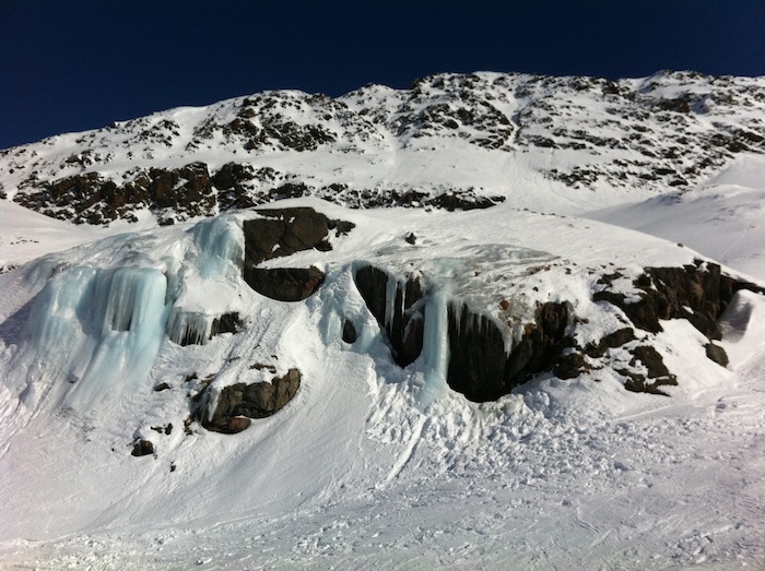
In March 2012 Chester Knee Clinic travelled to one of our favourite places, Kaunertal Glacier, for a bit of Spring skiing in Austria. Thanks to the resort's high altitude and the Kaunertal Glacier acting as a giant refrigerator, the snow conditions were fantastic despite the relatively warm and sunny weather. We all skied very well after our time in Jackson Hole and enjoyed traversing from the lifts to the vast areas of powder in the glacier's natural bowl. We love this small resort for its lack of crowds, beautiful scenery and guaranteed snow almost year-round.
We also ventured to the nearby resorts of Serfaus-Fiss-Ladis, which make up an immense ski area connected by an extensive
lift network. The views were breathtaking and the ski area provided and endless choice of slopes for all levels.
This page was launched on 1 March 2012. Last update: 12 October 2012.
Site last updated on: 28 March 2014
|
 Disclaimer: This website is a source of information
and education resource for health professionals and individuals
with knee problems. Neither Chester Knee Clinic nor Vladimir Bobic
make any warranties or guarantees that the information contained
herein is accurate or complete, and are not responsible for
any errors or omissions therein, or for the results obtained from
the use of such information. Users of this information are encouraged
to confirm the accuracy and applicability thereof with other sources.
Not all knee conditions and treatment modalities are described
on this website. The opinions and methods of diagnosis and treatment
change inevitably and rapidly as new information becomes available,
and therefore the information in this website does not necessarily
represent the most current thoughts or methods. The content of
this website is provided for information only and is not intended
to be used for diagnosis or treatment or as a substitute for consultation
with your own doctor or a specialist. Email
addresses supplied are provided for basic enquiries and should
not be used for urgent or emergency requests, treatment of any
knee injuries or conditions or to transmit confidential or medical
information. If you have sustained a knee injury or have a medical condition,
you should promptly seek appropriate medical advice from your local
doctor. Any opinions or information,
unless otherwise stated, are those of Vladimir Bobic, and in no
way claim to represent the views of any other medical professionals
or institutions, including Nuffield Health and Spire Hospitals. Chester
Knee Clinic will not be liable for any direct, indirect,
consequential, special, exemplary, or other damages, loss or injury
to persons which may occur by the user's reliance on any statements,
information or advice contained in this website. Chester Knee Clinic is
not responsible for the content of external websites.
Disclaimer: This website is a source of information
and education resource for health professionals and individuals
with knee problems. Neither Chester Knee Clinic nor Vladimir Bobic
make any warranties or guarantees that the information contained
herein is accurate or complete, and are not responsible for
any errors or omissions therein, or for the results obtained from
the use of such information. Users of this information are encouraged
to confirm the accuracy and applicability thereof with other sources.
Not all knee conditions and treatment modalities are described
on this website. The opinions and methods of diagnosis and treatment
change inevitably and rapidly as new information becomes available,
and therefore the information in this website does not necessarily
represent the most current thoughts or methods. The content of
this website is provided for information only and is not intended
to be used for diagnosis or treatment or as a substitute for consultation
with your own doctor or a specialist. Email
addresses supplied are provided for basic enquiries and should
not be used for urgent or emergency requests, treatment of any
knee injuries or conditions or to transmit confidential or medical
information. If you have sustained a knee injury or have a medical condition,
you should promptly seek appropriate medical advice from your local
doctor. Any opinions or information,
unless otherwise stated, are those of Vladimir Bobic, and in no
way claim to represent the views of any other medical professionals
or institutions, including Nuffield Health and Spire Hospitals. Chester
Knee Clinic will not be liable for any direct, indirect,
consequential, special, exemplary, or other damages, loss or injury
to persons which may occur by the user's reliance on any statements,
information or advice contained in this website. Chester Knee Clinic is
not responsible for the content of external websites.
[ back to top ]